Description
Elevate Your Diagnostics: Advanced Digital Dental X-Ray Systems
Revolutionize your dental practice with our state-of-the-art Digital Dental X-Ray Systems. Designed for precision, efficiency, and unparalleled patient safety, our range of imaging solutions provides the crystal-clear diagnostic insights you need to deliver superior patient care. From routine check-ups to complex treatment planning, our systems empower you with comprehensive visual data, transforming the way you practice dentistry.
Key Benefits for Your Practice:
- Superior Image Quality: Capture high-resolution images with exceptional clarity and detail, revealing even the most subtle pathologies.
- Reduced Radiation Dose: Protect your patients and staff with ultra-low dose technology, adhering to the highest safety standards.
- Enhanced Workflow Efficiency: Streamline your imaging process with intuitive software, rapid image acquisition, and instant access to digital files.
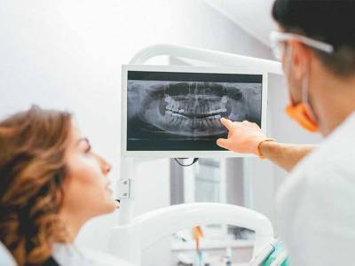
- Improved Patient Communication: Engage patients effectively with vivid visual aids, fostering better understanding and acceptance of treatment plans.
- Cost-Effective & Eco-Friendly: Eliminate chemical processing and film waste, reducing operational costs and your environmental footprint.
- Future-Proof Investment: Our systems are built with upgradeability and seamless integration in mind, adapting to the evolving needs of your practice.
Comprehensive Product Range:
We offer a diverse portfolio of digital X-ray solutions to meet the specific demands of any dental practice:
1. Intraoral X-Ray Units & Sensors/Plates
Our intraoral systems provide detailed views of individual teeth, root structures, and surrounding bone.
- High-Frequency X-Ray Generators:
- Advanced Control: Precise exposure settings (kVp, mA, exposure time) for optimal image density and contrast.
- Stable Output: Ensures consistent image quality, reducing retakes.
- Ergonomic Design: Available in wall-mounted, mobile, or handheld options for maximum flexibility and patient comfort.
- Small Focal Spot: Delivers razor-sharp images with minimal geometric distortion.
- Digital Intraoral Sensors:
- CMOS/CCD Technology: Available in various sizes (e.g., Size 1, Size 2) to fit diverse anatomies.
- Exceptional Durability: Robust design resistant to impact, bites, and liquids.
- Fast Acquisition: Instant image display, eliminating waiting times.
- Direct USB Connectivity: Plug-and-play simplicity.
- Phosphor Plate Systems (PSP):
- Film-Like Workflow: Ideal for practices transitioning from traditional film or preferring flexible sensor options.
- Thin & Flexible Plates: Patient-friendly and easy to position in challenging areas.
- High Resolution: Delivers excellent diagnostic quality.
- Reusable Plates: Cost-effective and environmentally friendly.
2. Panoramic X-Ray Systems
Capture a broad view of the entire dentition, temporomandibular joints (TMJ), and surrounding bone structures with a single exposure.
- 2D Panoramic Imaging:
- Crisp, Wide Views: Ideal for assessing impacted wisdom teeth, detecting cysts or tumors, evaluating periodontal disease, and pre-orthodontic assessment.
- Multi-Layer Technology: Some models offer multiple focal layers to compensate for slight positioning errors, ensuring optimal sharpness.
- Intuitive Positioning Aids: Laser guides and patient support systems ensure accurate and comfortable patient alignment.
- Cephalometric Options (Lateral Ceph, AP/PA Ceph):
- Dedicated Ceph Arm: For orthodontic analysis, growth studies, and surgical planning.
- Adjustable Field of View (FOV): Customize the imaging area based on diagnostic needs.
3. 3D Cone Beam Computed Tomography (CBCT)
For unparalleled diagnostic depth, our CBCT systems provide high-resolution volumetric images.
- Volumetric 3D Data: Offers detailed anatomical information from any angle, eliminating superimposition.
- Versatile Field of View (FOV): From small FOVs for single-tooth analysis to large FOVs encompassing the entire maxillofacial region.
- Clinical Applications:
- Implantology: Precise assessment of bone density and anatomy for accurate implant placement.
- Endodontics: Identification of accessory canals, root fractures, and periapical lesions.
- Oral Surgery: Impacted teeth, pathology detection, and surgical guide fabrication.
- Orthodontics: Airway analysis, TMJ evaluation, and complex treatment planning.
- Periodontics: Detailed visualization of bone loss and defects.
- Low Dose Protocols: Optimized for diagnostic efficacy while minimizing patient exposure.
Advanced Software Integration:
Our digital X-ray systems are complemented by powerful, user-friendly imaging software.
- Proprietary Imaging Software:
- Intuitive Interface: Easy navigation and image manipulation.
- Advanced Image Processing: Filters, sharpening, contrast adjustment, measurement tools, and density mapping.
- Diagnostic Tools: Caries detection, bone level analysis, endodontic measurement tools, implant planning modules.
- Patient Education Modules: Visually explain findings and treatment plans to patients.
- Seamless Integration:
- DICOM Compatibility: Ensures universal interoperability with third-party imaging software and PACS (Picture Archiving and Communication Systems).
- Practice Management Software (PMS) Integration: Direct access to patient images from your existing PMS for a streamlined workflow.
- AI-Powered Enhancements: (Optional) Advanced algorithms for automated caries detection, bone level measurement, and report generation, enhancing diagnostic confidence and efficiency.
Technical Specifications (General):
- Sensor Technology: CMOS/CCD (Intraoral), Flat Panel Detector (Panoramic/CBCT)
- Pixel Size: 20-100 µm (depending on model)
- Resolution: >10-25 lp/mm (depending on model)
- Tube Voltage (kVp): 60-90 kVp (adjustable)
- Tube Current (mA): 4-15 mA (adjustable)
- Exposure Time: 0.02 - 16 seconds (depending on acquisition type)
- Connectivity: USB 2.0/3.0, Ethernet
- Operating System Compatibility: Windows (specific versions), macOS (selected models)
- File Formats: DICOM, JPEG, TIFF, BMP
Why Choose Our Dental X-Ray Systems?
- Uncompromising Quality: Engineered with precision and robust materials for long-lasting performance and consistent image excellence.
- Commitment to Safety: Prioritizing patient and operator safety through advanced low-dose technology and rigorous quality control.
- Dedicated Support: Comprehensive installation, training, and ongoing technical support from our expert team.
- Innovation Leader: Continuously investing in research and development to bring you the latest advancements in dental imaging.
Ready to transform your diagnostic capabilities?
Request a Demo Today! | Download Our Brochure (PDF) | Visit Our Website: www.yourcompany.com
Contact Sales: Phone: [Your Phone Number] Email: [sales@yourcompany.com]