Description
This detailed product description is designed for a generic, high-quality 3D X-ray / Imaging system, emphasizing its benefits and applications across various medical and dental fields.
Unlocking New Dimensions in Diagnostics: Advanced 3D X-ray Imaging
In the evolving landscape of medical and dental diagnostics, traditional 2D X-rays, while invaluable, often present a limited, two-dimensional view, obscuring critical details due to superimposition. Enter the era of Advanced 3D X-ray Imaging, a revolutionary technology designed to provide clinicians with unprecedented insight into anatomical structures.
Our state-of-the-art 3D imaging system empowers practitioners to see beyond the conventional, offering a comprehensive, volumetric perspective that transforms diagnostic accuracy, treatment planning, and patient outcomes.
✨ Key Features That Redefine Clarity:
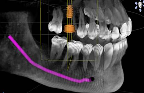
- Volumetric Precision: Captures comprehensive 3D data of the region of interest, eliminating distortion and superimposition inherent in 2D imaging. View anatomy from any angle, sliced in any plane.
- Exceptional Resolution: Delivers crystal-clear images with unparalleled detail, revealing intricate anatomical structures, subtle pathologies, and complex spatial relationships.
- Optimized Dose Protocols (Especially Cone Beam CT): Many systems offer significantly lower radiation exposure compared to conventional medical CT scans, enhancing patient safety while maintaining diagnostic quality.
- Rapid Acquisition: Swift scan times (typically 5-60 seconds) minimize patient discomfort, reduce motion artifacts, and streamline your workflow.
- Intuitive Software Suite: Advanced visualization and analysis software enables multi-planar reconstruction (MPR), 3D rendering, virtual surgical planning, precise measurements, and seamless integration with other digital tools.
- Flexible Field of View (FOV): Customizable imaging areas, from small, focused volumes for specific teeth or joints to larger volumes encompassing entire jaws, sinuses, or complex anatomical regions.
- Open Patient Positioning: Designed for patient comfort, often featuring open or wheelchair-accessible designs, accommodating a wide range of patient types and reducing claustrophobia.
💡 Transformative Benefits for Your Practice & Patients:
- Unrivaled Diagnostic Accuracy: Identify conditions, anomalies, and pathologies that might remain hidden or ambiguous on 2D images, leading to more confident and definitive diagnoses.
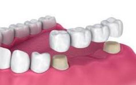
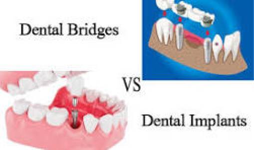
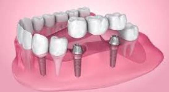
- Superior Treatment Planning: Confidently plan complex procedures, from surgical implant placement and orthodontic alignment to tumor localization, fracture repair, and airway assessment, with a complete understanding of the 3D anatomy.
- Enhanced Patient Communication: Engage patients with clear, easily understandable 3D visualizations of their anatomy, proposed treatments, and expected outcomes, fostering trust and compliance.
- Reduced Risk & Complications: Minimize surprises during surgery and improve the predictability of outcomes by thoroughly assessing anatomical variations and critical structures pre-operatively.
- Increased Efficiency & Workflow: Streamline your practice with faster diagnosis, more precise planning, and fewer necessary follow-up imaging appointments, saving valuable chair time for both staff and patients.
- Versatile Application: A single 3D scan provides a wealth of information for multiple diagnostic and planning needs, making it a powerful, multi-disciplinary tool.
🎯 Ideal for a Multitude of Clinical Applications:
Our 3D X-ray Imaging system is an indispensable tool across various specialties, including but not limited to:
- Dentistry & Oral Surgery:
- Dental implant planning and placement (bone quality, nerve proximity).
- Impacted wisdom teeth assessment (relationship to nerves, adjacent teeth).
- Orthodontic planning and airway analysis.
- Temporomandibular joint (TMJ) assessment.
- Endodontic evaluations (root canals, complex anatomy, fractures).
- Periodontal disease diagnosis and bone loss assessment.
- Oral pathology and lesion localization.
- ENT (Ear, Nose, Throat):
- Sinus analysis and pathology detection.
- Temporal bone imaging for hearing and balance disorders.
- Airway assessment for sleep apnea and other respiratory conditions.
- Foreign body localization.
- Orthopedics (Especially Extremities & Joints):
- Complex fracture evaluation and alignment.
- Joint analysis (degenerative changes, loose bodies).
- Pre-surgical planning for extremities.
- Maxillofacial Surgery:
- Trauma assessment and facial fracture planning.
- Tumor and cyst localization and staging.
- Reconstructive surgery planning.
- General Diagnostics:
- Wherever precise spatial relationships, volumetric data, and high-resolution imaging are critical for confident diagnosis and treatment.
🌐 Technical Specifications (Illustrative - Varies by System):
- Imaging Modality: Cone Beam Computed Tomography (CBCT) or Multi-Slice CT (depending on model)
- Detector Type: High-sensitivity Flat Panel Detector
- Field of View (FOV): Customizable; e.g., Ø5x5 cm to Ø20x20 cm
- Voxel Size: Down to 70-100 microns for ultra-high resolution
- Scan Time: Typically 5-60 seconds (depending on FOV and resolution setting)
- Radiation Dose: Optimized protocols for lowest possible dose, often significantly lower than conventional CT
- Image Reconstruction: Multi-planar (axial, coronal, sagittal) and 3D volumetric rendering
- Software Integration: DICOM 3.0 compatible, open architecture for seamless integration with various third-party planning and CAD/CAM software
- Power Requirements: Standard clinic/hospital electrical supply
- Footprint: Compact design for integration into various clinical settings
📈 Elevate Your Diagnostic Capabilities Today.
Investing in Advanced 3D X-ray Imaging isn't just an upgrade; it's a paradigm shift in patient care and diagnostic capability. Empower your practice with the tools to see more, diagnose with greater confidence, and deliver superior, safer treatment outcomes. Stay at the forefront of medical and dental innovation.
Ready to experience the clarity and confidence of 3D imaging? Contact us for a personalized demonstration or to learn how this advanced technology can transform your practice.