Description
Endoscopic Tympanoplasty
Minimally Invasive | Enhanced Visualization | Superior Patient Outcomes
Product Overview
Endoscopic Tympanoplasty represents a paradigm shift in the surgical repair of tympanic membrane perforations (eardrum holes) and certain middle ear pathologies. By utilizing advanced high-definition endoscopes, this innovative technique offers otolaryngologists an unparalleled, magnified, and panoramic view of the entire external auditory canal and middle ear space. It facilitates the precise repair of the tympanic membrane and associated structures entirely through the natural ear canal, often eliminating the need for traditional external incisions and extensive tissue dissection associated with microscopic approaches.
This minimally invasive procedure is designed to restore hearing, prevent recurrent infections, and improve the overall quality of life for patients suffering from chronic eardrum perforations.
Key Features & Advantages
- Minimally Invasive Approach: Performed entirely through the external auditory canal, precluding the need for post-auricular (behind-the-ear) or endaural incisions in most cases. This reduces surgical trauma and preserves external ear anatomy.
- Superior Endoscopic Visualization: Provides a wide-field, high-definition, and brightly illuminated view of the tympanic membrane and middle ear structures. Endoscopes (0-degree and angled options) allow for direct visualization of anterior and inferior recesses, which are often blind spots or require extensive drilling with microscopic techniques.
- Enhanced Surgical Access: The angulated optics and slender profile of the endoscope offer unparalleled access to challenging areas within the middle ear, improving the efficiency and precision of graft placement and ossicular manipulation.
- Reduced Patient Morbidity: Leads to significantly less post-operative pain, swelling, and discomfort compared to traditional open surgery.
- Faster Recovery Times: Patients typically experience a quicker return to normal activities, with less need for strong pain medication.
- Improved Cosmetic Outcomes: Absence of external incisions results in no visible scars, a significant benefit for patient satisfaction.
- Potential for Shorter Operating Room Time: For experienced endoscopic surgeons, the direct visualization and streamlined approach can lead to reduced procedural duration.
- Facilitates Intraoperative Teaching: The video feed from the endoscope can be displayed on a large monitor, making it an excellent tool for surgical training and observation.
Indications
Endoscopic Tympanoplasty is indicated for the repair of a variety of middle ear conditions, including:
- Tympanic Membrane Perforations: Small to medium-sized central or marginal perforations resulting from infection, trauma, or previous surgery.
- Retraction Pockets: Repair of deep retraction pockets without significant cholesteatoma formation.
- Myringoplasty: Grafting procedures to reconstruct the tympanic membrane.
- Ossicular Chain Discontinuity: Repair of disruptions to the small bones of hearing (ossicles), such as incus long process erosion, when feasible endoscopically.
- Combined Procedures: Can be integrated with other minimally invasive middle ear explorations or minor ossiculoplasties.
Procedure Overview (General)
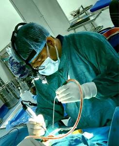
The procedure is typically performed under local anesthesia with sedation or general anesthesia. A rigid, high-definition endoscope is carefully advanced into the external auditory canal, providing the surgeon with a magnified, comprehensive view of the perforation. The edges of the perforation are precisely denuded, and a graft (commonly temporalis fascia, tragal perichondrium, or cartilage) is harvested and prepared. The graft is then meticulously positioned, usually in an underlay or overlay technique, entirely endoscopically, to reconstruct the tympanic membrane. Packing material may be placed in the ear canal to stabilize the graft.
Target Audience
- Otolaryngologists (ENT Surgeons): Seeking to offer cutting-edge, patient-friendly, and effective solutions for tympanic membrane repair.
- Surgical Centers & Hospitals: Aiming to enhance their otologic surgery offerings with modern, minimally invasive techniques that improve patient satisfaction and surgical efficiency.
- Fellows & Residents in Otology: An excellent platform for learning middle ear anatomy and surgical techniques due to the direct, magnified visualization.
Benefits for the Surgeon & Practice
- Expanded Surgical Capabilities: Positions your practice at the forefront of otologic innovation.
- Increased Patient Referrals: Improved patient satisfaction due to less pain, faster recovery, and no visible scarring leads to positive word-of-mouth and referrals.
- Streamlined Workflow: Reduced need for complex mastoidectomy setups or extensive drilling in select cases.
- Enhanced Precision: The high-resolution, magnified view allows for meticulous tissue manipulation and graft placement.
- Competitive Advantage: Differentiates your practice by offering a less invasive alternative to traditional tympanoplasty.
Essential Equipment
- High-Definition Rigid Otologic Endoscopes: Typically 2.7mm or 3.0mm diameter, with 0-degree and 30-degree angulations for comprehensive viewing.
- High-Definition Camera System & Monitor: For clear visualization and recording.
- Fiber Optic Light Source: Powerful and reliable illumination.
- Specialized Endoscopic Otologic Instruments: Longer, thinner instruments designed to work alongside the endoscope, including picks, dissectors, scissors, and cup forceps.
- Standard Graft Harvesting Instruments: For collecting temporalis fascia, tragal perichondrium, or cartilage.
- Suction & Irrigation Capabilities.
Outcomes & Efficacy
Clinical studies and extensive surgical experience have demonstrated that Endoscopic Tympanoplasty achieves comparable or often superior graft take rates to microscopic techniques, while offering significant advantages in terms of reduced invasiveness and patient comfort. Success rates typically range from 85-95%, influenced by factors such as perforation size, location, underlying ear disease, and surgeon expertise.
Why Choose Endoscopic Tympanoplasty?
Embrace the evolution of otologic surgery. Endoscopic Tympanoplasty provides a sophisticated, patient-centric approach to middle ear repair, delivering excellent functional outcomes with significantly reduced post-operative morbidity. It's not merely a procedural change; it's a commitment to advanced patient care, surgical precision, and a superior recovery experience.
Further Information: For detailed technical specifications, training opportunities, or to discuss how to integrate Endoscopic Tympanoplasty into your surgical practice, please contact our surgical innovations team.
Disclaimer: All surgical procedures carry inherent risks. Patient selection, surgeon experience, and proper technique are crucial for optimal outcomes. This information is intended for professional medical audiences and should not be used as a substitute for professional medical advice.